© Didier MONTARRAS/Institut Pasteur/CNRS Images
Reference
20130001_1641
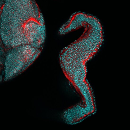
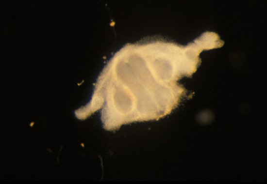
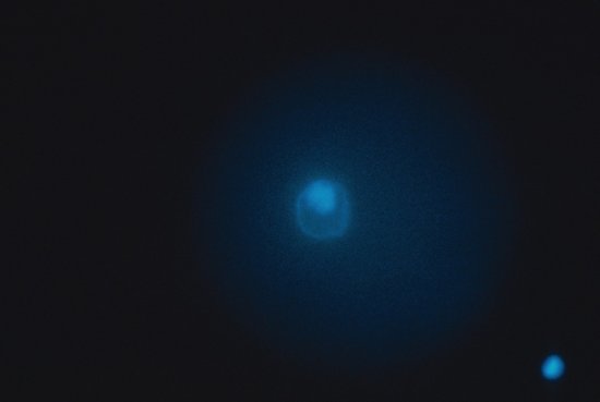
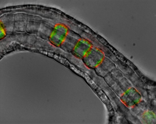
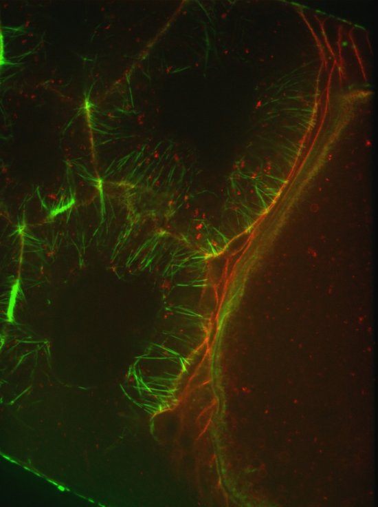
Transverse section of the muscle of a mouse
Transverse section of the muscle of a mouse. On the left, the green staining reveals the expression of the GFP protein (Green Fluorescent Protein), under the control of the Pax3 gene, in the nucleus of a myosatellite cell. On the right, the blue staining marks all the nuclei. The red staining, on both images, marks the presence of the extracellular matrix protein, laminin, around the fibers. The capacity of skeletal muscles to undergo regeneration depends on skeletal muscle stem cells, called myosatellite cells. Upon muscle injury, myosatellite cells undergo activation, proliferate, differentiate and fuse with each other to form new fibers.
Add to my selection
Terms of use
The use of media visible on the CNRS Images Platform can be granted on request. Any reproduction or representation is forbidden without prior authorization from CNRS Images (except for resources under Creative Commons license).
No modification of an image may be made without the prior consent of CNRS Images.
No use of an image for advertising purposes or distribution to a third party may be made without the prior agreement of CNRS Images.
For more information, please consult our general conditions